Professor Nigel W. John FEG FLSW
Home Research About Contact
3D Ultrasound (1995-7)
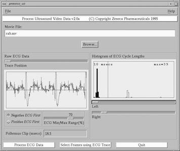 The GUI for the software that processed the Ultrasound data.
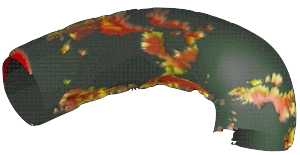
The most enjoyable project when working for Zeneca Pharmaceuticals was to be part of the team that developed the world’s first system for reonstructing 3D Ultrasound data from freehand 2D ultrasound images (Barry et al., 1997). The goal was to measure plaque build up on the artery wall of watanabe rabbits to measure if the drugs in development would reduce the amount of plaque - see image above. The innovation was to attach an electromagnetic position tracker to the ultrasound transducer so that we knew the position and orientation of each 2D ultrasound image. We also recorded the ECG signal so that the 3D reconstruction only used images from when the heart is at rest (diastole). I wrote all of the software for processing the data ready for reconstruction into a voxel data set.
The GUI for the software that processed the Ultrasound data.
The most enjoyable project when working for Zeneca Pharmaceuticals was to be part of the team that developed the world’s first system for reonstructing 3D Ultrasound data from freehand 2D ultrasound images (Barry et al., 1997). The goal was to measure plaque build up on the artery wall of watanabe rabbits to measure if the drugs in development would reduce the amount of plaque - see image above. The innovation was to attach an electromagnetic position tracker to the ultrasound transducer so that we knew the position and orientation of each 2D ultrasound image. We also recorded the ECG signal so that the 3D reconstruction only used images from when the heart is at rest (diastole). I wrote all of the software for processing the data ready for reconstruction into a voxel data set.
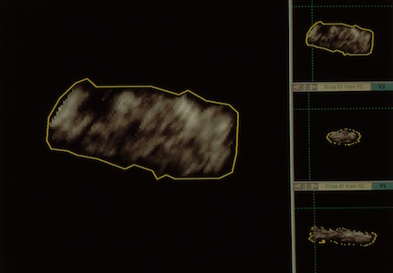 TOSCA (Tool for Segmentation, Correlation and Analysis).
We used software from IBM called TOSCA to segment the voxel data, and IBM’s Data Explorer tool for visualisation.
TOSCA (Tool for Segmentation, Correlation and Analysis).
We used software from IBM called TOSCA to segment the voxel data, and IBM’s Data Explorer tool for visualisation.
Further Reading
- Barry, C. D., Allott, C. P., John, N. W., Mellor, P. M., Arundel, P. A., Thomson, D. S., & Waterton, J. C. (1997). Three-dimensional freehand ultrasound: Image reconstruction and volume analysis. Ultrasound in Medicine and Biology, 23(8), 1209–1224.